Electron microscopy
The Centre for Environmental Scanning Electron Microscopy (CESEM) was set up in 1998, to facilitate state-of-the-art research within the Institute of GeoEnergy Engineering (IGE) and foster collaboration across Heriot-Watt University. The facility is accessible to all students and staff across the University, and is also available for external commercial users.
Equipment
The Centre currently has two scanning electron microscopes: an XL30 ESEM with LaB6 filament, and a Quanta 650 FEG SEM. Both are equipped with a range of detectors and stages, including energy dispersive x-ray (EDX) capabilities, and are ideally suited for a wide range of investigations.
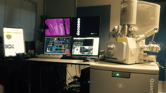
Quanta FEG 650 Suite
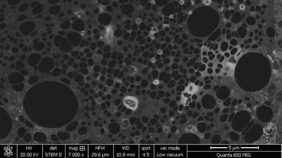
Quanta FEG 650 SEM equipped with Oxford Instruments X-maxN 150 EDX detector. Also equipped with a range of secondary (SE, GSE) and backscattered (BSE) detectors, a cathodoluminescence (CL) detector, an electron back scattered diffraction (EBSD) detector, and a wet scanning transmission electron microscopy (STEM) detector.
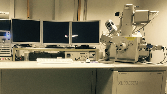
XL30 LaB6 ESEM Suite
XL30 LaB6 ESEM equipped with Oxford Instruments X-max 80 EDX detector. Also equipped with a range of secondary (SE, GSE) detectors, a backscattered (BSE) detector, and a cathodoluminescence (CL) detector. This suite also has a range of additional stages: Peltier cooling stage; thousand degree heating stage; two tensile-compression stages; microinjector; and cryo-stage (currently not in use).
Modes of Operation
| Mode | Purpose |
|---|---|
| High vacuum | Used for electrically conductive stable samples, such as metals, and non-conductive samples coated with gold or carbon. Good for standard SEM and EDX analysis of a wide range of materials. |
| Low vacuum | For clean uncoated non-conductive samples: ceramics, glass, plastics and resins, geological materials, cements and concrete, filter papers, pharmaceuticals, paper, chocolate etc. Typically operated at 0.8 Torr, with a water vapour atmosphere. |
| Full ESEM (wet mode) | This is typically used for wet or oily non-vacuum compatible samples: hydrated clays, emulsions, medical materials, micro-organisms, aphids etc. In Full ESEM mode, the chamber is held at between 2-6 Torr, and samples are cooled to 5˚C. Water vapour (H2O) is used as an imaging gas, to promote charge suppression, and maintain sample humidity. Where there is no requirement to maintain hydrated samples, other gases such as carbon dioxide or nitrogen can be used instead. |
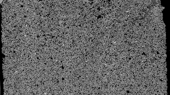
Large format high-resolution imaging
Using ‘MAPS' software, images can be automatically collected and stitched together (fig. 3), with the construction of montaged images comprising several thousand tiled frames being easily achieved. This allows high-resolution images to be collected over relatively large areas (several square centimetres), such images can be reviewed at a later date in a fashion akin to having a virtual microscope on your own desktop.
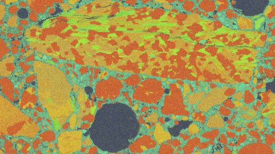
Elemental mapping
‘Aztec Large Area Maps' software can be used for the automated capture and stitching of elemental composition maps, across areas of several square centimetres, or individual frames. Once collected, individual maps can be montaged and exported as large format maps. Individual tiles and montaged maps can be manipulated to emphasis features, and both qualitative (spectrum) and quantitative data can be extracted from selected areas.
Particle analysis
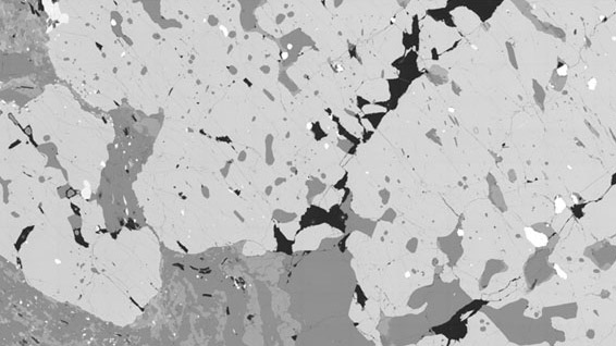
The programmes ‘IncaFeature' and ‘AZtecFeature' can be used for the identification and analysis of particulate materials. Particularly suitable for heavy mineral analysis, the analysis of industrial heavy metal contamination, and the analysis of particulate materials on filter papers. Thresholding and segmentation of backscattered (BSE) images can be used to select phases of interest for analysis. Data can be collected on size, shape, orientation, position and elemental composition, which can be exported in the form of an Excel file, for plotting using third party software.
EBSD analysis
The EBSD detector can be used to collect data on phase distribution and crystal orientation. Samples need to be flat, and normally require chemical-mechanical polishing, or broad beam ion-milling, to limit surface damage within the top 100 nm or so of the surface. EBSD maps can be collected using the ‘Aztec' EBSD mapping software, including the large area mapping (LAM) function.
Dynamic observations
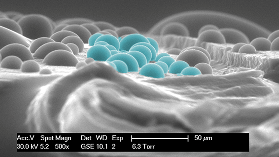
The ESEM chamber can be thought of as a dynamic experimental chamber, in which a variety of experiments can be observed at a micron level. Examples include wetting and drying, wettability observations, freeze – thaw cycling, heating and melting, salt crystallization, and compressive or tensile loading. The ESEM is particularly useful for performing wettability observations at the micron level, making it possible to quantify differences in wettability at the pore scale - see https://dx.doi.org/10.13140/RG.2.1.1813.2246.
Bookings
We regularly provide consultancy services to the oil industry, as well as to the pharmaceuticals and electronics industries, offering a fast turnaround at a competitive price. All enquiries and requests for bookings should be made via e-mail to the facility manager Dr Jim Buckman.
Wettability experiment inside an ESEM (Quanta 650 FEG SEM), showing three water wetting and drying cycles of micro glass beads. The beads are hydrophilic, therefore water droplets with a contact angle of less than 90 degrees can be seen forming on the surface. Because of the hydrophilic nature, water bridges between grains form and are maintained, but expand and contract during wetting and drying cycles.
Glauconite grain, Oligocene, New Zealand. A wettability experiment inside an ESEM chamber (Quanta 650 FEG SEM). Six wetting and drying cycles of highly hydrophilic glauconite. The glauconite is highly hydrophilic, to the extent that condensing water is initially absorbed by the grain (although no swelling observed), and no water droplets are observed on the grain surface. Then water starts to rise along the cracks, before the whole surface is overwhelmed. As the surface floods with water, coccolith micro fossils can be seen floating around, which are presumably more hydrophobic and repelled from the water.
Selected Specialist Capabilities

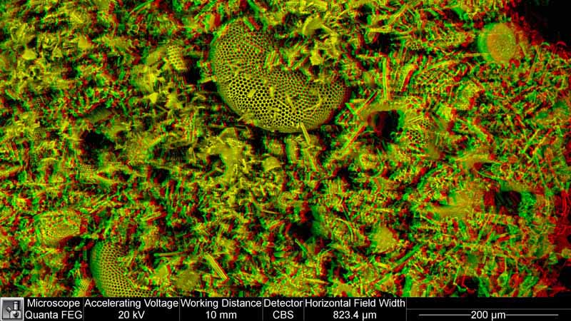
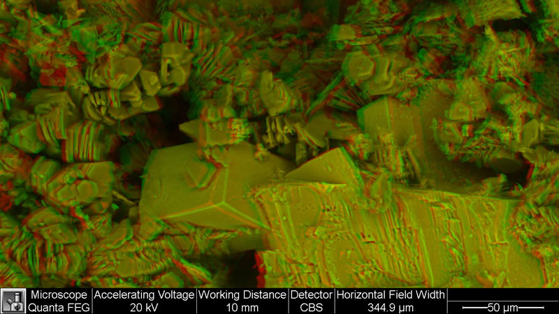
Stereo Anaglyphs
Key information
Dr Jim Buckman
- Research Fellow
- j.buckman@hw.ac.uk